四篇綜述的詳細內容
選讀・IP 11・影像分割(二):主動輪廓——蛇模型與水平集 的論文導讀(第 1/2 篇)。引用編號對應本頁底部的參考文獻。
導覽:本頁與本系列: 導覽:本頁與本系列
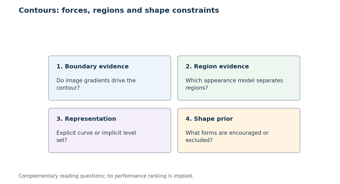
先辨認輪廓依賴邊界、區域外觀、拓樸表示,還是形狀先驗,再選擇對應回顧。
以下四篇綜述描繪了主動輪廓的全貌,以及它與深度學習的關係。
醫學影像中的可變形模型
McInerney 與 Terzopoulos [13] 寫下了醫學影像分析中可變形模型的經典早期綜述。他們把蛇模型及其 3D 延伸視為最小化能量的物理模型,並回顧它們在 CT、MRI 等模態上用於分割、形狀表示、比對與運動追蹤的應用。他們的主要結論至今仍成立:當影像本身含糊不清、需要模型提供平滑性與先驗知識時,可變形模型最有用。
統計式區域水平集
Cremers、Rousson 與 Deriche [14] 回顧以區域為主的水平集方法,並從同一個貝氏框架推導出它們:每個區域都有一個機率模型(針對亮度、色彩、紋理或運動),水平集要最大化這個分割的後驗機率,並可選擇性地加入統計形狀先驗。他們主張區域式能量比邊緣式蛇模型對雜訊較不敏感、局部最小值也較少,這正是後來的研究以 Chan–Vese 類型項為主流的原因。
主動輪廓模型的近期分類
Chen、Ge、Wang 與 Chen [15] 把主動輪廓模型分為邊緣式(GAC、DRLSE)、區域式(Chan–Vese,以及為處理亮度不均而設計的局部擬合變體)與混合式模型,實作了一組代表性模型,並在合成、醫學與自然影像上與深度學習分割比較。他們的結論很務實:傳統模型不需要訓練資料、能給出平滑的封閉邊界,但對初始化和參數敏感;在有標註資料的情況下,深度模型更準確;兩者是互補的。
深度分割中的形狀限制
Bohlender、Oksuz 與 Mukhopadhyay [16] 綜述了如何把解剖形狀知識注入深度醫學影像分割:條件隨機場、統計形狀模型、主動輪廓,以及符號距離圖等隱式形狀表示。他們的動機正是主動輪廓當初要修補的弱點:逐像素分類器可能產生破碎的區域與拓撲錯誤。
如何判讀這些論文的證據
應依模型相信哪些證據、能表示哪些形狀比較。邊緣輪廓依賴邊界,區域模型依賴外觀假設,水平集影響拓樸如何演變,形狀先驗則限制可接受解;這些選擇可同時存在,不必硬分成互斥類別。
除了重疊分數,也要檢查初始化、參數選擇與停止條件。形狀約束可排除不合理結果,也可能排除有效病灶或罕見解剖,屬於建模取捨。先讀綜述揭露的假設,再回原研究確認影像、前處理與監督是否一致;跨研究並列分數不能單獨歸因於輪廓能量。
參考文獻
- R. C. Gonzalez and R. E. Woods, Digital Image Processing, 4th ed., Pearson, 2018, Ch. 11. publisher page
- M. Kass, A. Witkin, and D. Terzopoulos, “Snakes: Active contour models,” International Journal of Computer Vision, vol. 1, no. 4, pp. 321–331, 1988. doi:10.1007/BF00133570
- C. Xu and J. L. Prince, “Snakes, shapes, and gradient vector flow,” IEEE Transactions on Image Processing, vol. 7, no. 3, pp. 359–369, 1998; see also C. Xu, A. Yezzi Jr., and J. L. Prince, “On the relationship between parametric and geometric active contours,” Proc. 34th Asilomar Conf. on Signals, Systems, and Computers, 2000. GVF project page with both papers
- L. D. Cohen, “On active contour models and balloons,” CVGIP: Image Understanding, vol. 53, no. 2, pp. 211–218, 1991. author PDF
- S. Osher and J. A. Sethian, “Fronts propagating with curvature-dependent speed: Algorithms based on Hamilton–Jacobi formulations,” Journal of Computational Physics, vol. 79, pp. 12–49, 1988. doi:10.1016/0021-9991(88)90002-2
- V. Caselles, R. Kimmel, and G. Sapiro, “Geodesic active contours,” International Journal of Computer Vision, vol. 22, no. 1, pp. 61–79, 1997. PDF
- D. Mumford and J. Shah, “Optimal approximations by piecewise smooth functions and associated variational problems,” Communications on Pure and Applied Mathematics, vol. 42, no. 5, pp. 577–685, 1989. doi:10.1002/cpa.3160420503
- T. F. Chan and L. A. Vese, “Active contours without edges,” IEEE Transactions on Image Processing, vol. 10, no. 2, pp. 266–277, 2001. doi:10.1109/83.902291
- D. Adalsteinsson and J. A. Sethian, “A fast level set method for propagating interfaces,” Journal of Computational Physics, vol. 118, pp. 269–277, 1995. summary page
- C. Li, C. Xu, C. Gui, and M. D. Fox, “Distance regularized level set evolution and its application to image segmentation,” IEEE Transactions on Image Processing, vol. 19, no. 12, pp. 3243–3254, 2010. doi:10.1109/TIP.2010.2069690
- P. Márquez-Neila, L. Baumela, and L. Álvarez, “A morphological approach to curvature-based evolution of curves and surfaces,” IEEE Transactions on Pattern Analysis and Machine Intelligence, 2014. doi:10.1109/TPAMI.2013.106
- scikit-image developers, “skimage.segmentation” API reference (
active_contour,chan_vese,morphological_chan_vese,morphological_geodesic_active_contour). docs - T. McInerney and D. Terzopoulos, “Deformable models in medical image analysis: A survey,” Medical Image Analysis, vol. 1, no. 2, 1996. author PDF
- D. Cremers, M. Rousson, and R. Deriche, “A review of statistical approaches to level set segmentation: Integrating color, texture, motion and shape,” International Journal of Computer Vision, vol. 72, no. 2, pp. 195–215, 2007. doi:10.1007/s11263-006-8711-1
- Y. Chen, P. Ge, G. Wang, and H. Chen, “An overview of intelligent image segmentation using active contour models,” Intelligence & Robotics, vol. 3, no. 1, pp. 23–55, 2023. open access
- S. Bohlender, I. Oksuz, and A. Mukhopadhyay, “A survey on shape-constraint deep learning for medical image segmentation,” IEEE Reviews in Biomedical Engineering, vol. 16, pp. 225–240, 2023. arXiv:2101.07721
- S. Peng, W. Jiang, H. Pi, X. Li, H. Bao, and X. Zhou, “Deep snake for real-time instance segmentation,” CVPR, 2020. arXiv:2001.01629
- X. Chen, B. M. Williams, S. R. Vallabhaneni, G. Czanner, R. Williams, and Y. Zheng, “Learning active contour models for medical image segmentation,” CVPR, 2019. CVF open access
- B. Kim and J. C. Ye, “Mumford–Shah loss functional for image segmentation with deep learning,” IEEE Transactions on Image Processing, vol. 29, pp. 1856–1866, 2020. doi · arXiv:1904.02872
- H. Kervadec, J. Bouchtiba, C. Desrosiers, E. Granger, J. Dolz, and I. Ben Ayed, “Boundary loss for highly unbalanced segmentation,” Medical Image Analysis, vol. 67, 2021 (first presented at MIDL 2019). arXiv:1812.07032