The four surveys in more detail
Optional · paper guide 1 of 2 for IP 11 · Image Segmentation II: Active Contours — Snakes and Level Sets. Citation numbers refer to the reference list at the bottom of this page.
Navigate: this page and this track: Navigate: this page and this track
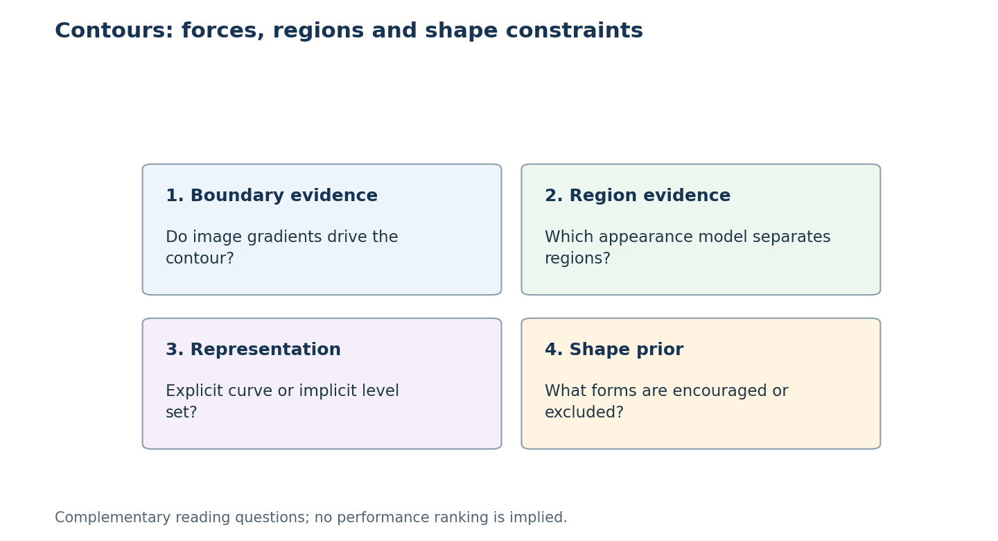
Choose a reading by topic: Boundary evidence, Region evidence, Representation, Shape prior.
Four surveys map active contours and their place next to deep learning.
Deformable models in medical imaging
McInerney and Terzopoulos [13] wrote the standard early survey of deformable models in medical image analysis. They frame snakes and their 3D relatives as energy-minimizing physical models and review applications to segmentation, shape representation, matching and motion tracking across CT, MRI and other modalities. Their main takeaway still holds: deformable models are most useful when the image alone is ambiguous and the model must contribute smoothness and prior knowledge.
Statistical region-based level sets
Cremers, Rousson and Deriche [14] review region-based level-set methods and derive them from a common Bayesian framework: each region gets a probability model (for intensity, color, texture or motion), and the level set maximizes the posterior probability of the partition, optionally with a statistical shape prior. Their argument for region-based energies, which are less sensitive to noise and have fewer local minima than edge-based snakes, is the reason Chan–Vese-type terms dominate later work.
A recent taxonomy of active contour models
Chen, Ge, Wang and Chen [15] sort active contour models into edge-based (GAC, DRLSE), region-based (Chan–Vese and local-fitting variants designed for intensity inhomogeneity) and hybrid models, implement a set of representative models, and compare them with deep learning segmentation on synthetic, medical and natural images. Their conclusion is pragmatic: classical models need no training data and give smooth closed boundaries, but they are sensitive to initialization and parameters; deep models are more accurate where labelled data exist; the two are complementary.
Shape constraints in deep segmentation
Bohlender, Oksuz and Mukhopadhyay [16] survey how anatomical shape knowledge is injected into deep medical image segmentation: conditional random fields, statistical shape models, active contours, and implicit shape representations such as signed distance maps. Their motivation is exactly the weakness active contours were designed to fix: pixel-wise classifiers can produce fragmented regions and topological errors.
Reading these papers critically
Compare deformable models by the evidence they trust and the shapes they can represent. An edge-driven contour needs useful boundary evidence; a region model needs an appearance assumption; a level-set representation changes how topology can evolve; and a shape constraint restricts the set of acceptable solutions. These are partly independent choices. A method can use a region term and a shape prior without belonging to only one survey category.
For a segmentation result, inspect initialization, parameter selection and stopping as well as overlap scores. Sensitivity to a starting curve can matter more in use than one favorable average. Shape constraints can reject implausible predictions but also exclude valid pathology or unusual anatomy; that is a modeling tradeoff, not automatically an improvement. Read survey comparisons for the assumptions they expose, then use original studies to establish whether the same images, preprocessing and supervision were used. Numerical tables assembled from separate studies do not isolate the contour energy as the sole cause of a difference.
References
- R. C. Gonzalez and R. E. Woods, Digital Image Processing, 4th ed., Pearson, 2018, Ch. 11. publisher page
- M. Kass, A. Witkin, and D. Terzopoulos, “Snakes: Active contour models,” International Journal of Computer Vision, vol. 1, no. 4, pp. 321–331, 1988. doi:10.1007/BF00133570
- C. Xu and J. L. Prince, “Snakes, shapes, and gradient vector flow,” IEEE Transactions on Image Processing, vol. 7, no. 3, pp. 359–369, 1998; see also C. Xu, A. Yezzi Jr., and J. L. Prince, “On the relationship between parametric and geometric active contours,” Proc. 34th Asilomar Conf. on Signals, Systems, and Computers, 2000. GVF project page with both papers
- L. D. Cohen, “On active contour models and balloons,” CVGIP: Image Understanding, vol. 53, no. 2, pp. 211–218, 1991. author PDF
- S. Osher and J. A. Sethian, “Fronts propagating with curvature-dependent speed: Algorithms based on Hamilton–Jacobi formulations,” Journal of Computational Physics, vol. 79, pp. 12–49, 1988. doi:10.1016/0021-9991(88)90002-2
- V. Caselles, R. Kimmel, and G. Sapiro, “Geodesic active contours,” International Journal of Computer Vision, vol. 22, no. 1, pp. 61–79, 1997. PDF
- D. Mumford and J. Shah, “Optimal approximations by piecewise smooth functions and associated variational problems,” Communications on Pure and Applied Mathematics, vol. 42, no. 5, pp. 577–685, 1989. doi:10.1002/cpa.3160420503
- T. F. Chan and L. A. Vese, “Active contours without edges,” IEEE Transactions on Image Processing, vol. 10, no. 2, pp. 266–277, 2001. doi:10.1109/83.902291
- D. Adalsteinsson and J. A. Sethian, “A fast level set method for propagating interfaces,” Journal of Computational Physics, vol. 118, pp. 269–277, 1995. summary page
- C. Li, C. Xu, C. Gui, and M. D. Fox, “Distance regularized level set evolution and its application to image segmentation,” IEEE Transactions on Image Processing, vol. 19, no. 12, pp. 3243–3254, 2010. doi:10.1109/TIP.2010.2069690
- P. Márquez-Neila, L. Baumela, and L. Álvarez, “A morphological approach to curvature-based evolution of curves and surfaces,” IEEE Transactions on Pattern Analysis and Machine Intelligence, 2014. doi:10.1109/TPAMI.2013.106
- scikit-image developers, “skimage.segmentation” API reference (
active_contour,chan_vese,morphological_chan_vese,morphological_geodesic_active_contour). docs - T. McInerney and D. Terzopoulos, “Deformable models in medical image analysis: A survey,” Medical Image Analysis, vol. 1, no. 2, 1996. author PDF
- D. Cremers, M. Rousson, and R. Deriche, “A review of statistical approaches to level set segmentation: Integrating color, texture, motion and shape,” International Journal of Computer Vision, vol. 72, no. 2, pp. 195–215, 2007. doi:10.1007/s11263-006-8711-1
- Y. Chen, P. Ge, G. Wang, and H. Chen, “An overview of intelligent image segmentation using active contour models,” Intelligence & Robotics, vol. 3, no. 1, pp. 23–55, 2023. open access
- S. Bohlender, I. Oksuz, and A. Mukhopadhyay, “A survey on shape-constraint deep learning for medical image segmentation,” IEEE Reviews in Biomedical Engineering, vol. 16, pp. 225–240, 2023. arXiv:2101.07721
- S. Peng, W. Jiang, H. Pi, X. Li, H. Bao, and X. Zhou, “Deep snake for real-time instance segmentation,” CVPR, 2020. arXiv:2001.01629
- X. Chen, B. M. Williams, S. R. Vallabhaneni, G. Czanner, R. Williams, and Y. Zheng, “Learning active contour models for medical image segmentation,” CVPR, 2019. CVF open access
- B. Kim and J. C. Ye, “Mumford–Shah loss functional for image segmentation with deep learning,” IEEE Transactions on Image Processing, vol. 29, pp. 1856–1866, 2020. doi · arXiv:1904.02872
- H. Kervadec, J. Bouchtiba, C. Desrosiers, E. Granger, J. Dolz, and I. Ben Ayed, “Boundary loss for highly unbalanced segmentation,” Medical Image Analysis, vol. 67, 2021 (first presented at MIDL 2019). arXiv:1812.07032